How Ultrasound Scans Help Diagnose Internal Organ and Tissue Conditions
Ultrasound scanning is one of the most widely used and reliable diagnostic tools in modern medicine. It provides a safe, non-invasive, and painless way to visualize internal organs, tissues, and blood flow in real time. From evaluating abdominal pain to monitoring pregnancy, ultrasound plays an essential role in early diagnosis and treatment planning.
An Ultrasonogrāfija (USG) is a medical imaging technique that uses high-frequency sound waves to produce detailed images of the body’s internal structures. Unlike X-rays or CT scans, ultrasound does not use ionizing radiation, making it safe for patients of all ages, including pregnant women and children. This imaging method helps doctors assess organ health, detect abnormalities, and guide medical procedures with precision.
What Is Ultrasound and How Does It Work?
Ultrasound imaging works by emitting sound waves through a handheld device called a transducer. When these sound waves bounce off tissues, organs, and fluids inside the body, they create echoes that are converted into live images on a monitor. The procedure is painless, quick, and typically takes between 15 and 45 minutes, depending on the area being examined.
Ultrasound can be performed externally on the skin’s surface or internally through specialized probes, depending on the part of the body being evaluated. It is commonly used for diagnostic purposes, monitoring, and image-guided treatments.
Common Uses of Ultrasound Scans
Ultrasound scans are used to evaluate a wide range of medical conditions. Some of the most common applications include:
1. Abdominal Ultrasound: Used to examine organs such as the liver, kidneys, gallbladder, pancreas, and spleen. It helps detect gallstones, cysts, or liver diseases.
2. Pelvic Ultrasound: Commonly performed to assess the uterus, ovaries, and bladder in women, and the prostate gland in men.
3. Obstetric Ultrasound: Monitors fetal development during pregnancy, checks for complications, and estimates gestational age.
4. Vascular Ultrasound: Evaluates blood flow in arteries and veins to detect blockages, clots, or circulatory problems.
5. Thyroid and Neck Ultrasound: Used to examine the thyroid gland and identify nodules or cysts.
6. Musculoskeletal Ultrasound: Helps diagnose soft tissue injuries, tendon tears, or joint inflammation.
These versatile applications make ultrasound an essential diagnostic tool across multiple medical specialties.
Advantages of Ultrasound Imaging
Ultrasound offers several benefits over other imaging techniques. It is safe, as it does not expose patients to radiation, and provides real-time imaging that allows doctors to observe organ movement, blood flow, and tissue structure dynamically.
Another advantage is its accessibility and affordability. Ultrasound machines are portable and can be used in hospitals, clinics, or even bedside settings. Additionally, the results are available immediately, enabling quick diagnosis and treatment decisions.
Ultrasound is also widely used for guiding medical procedures such as biopsies, fluid drainage, and injections, increasing accuracy and minimizing risks.
Detecting and Monitoring Internal Organ Conditions
Ultrasound plays a critical role in detecting abnormalities in organs and soft tissues. It can identify cysts, tumors, inflammation, infections, and structural abnormalities. For example, in liver evaluations, ultrasound helps detect fatty liver disease, cirrhosis, or tumors. In the kidneys, it can identify stones, infections, or obstructions.
For heart and vascular conditions, Doppler ultrasound measures blood flow and detects issues such as arterial blockages or blood clots. In musculoskeletal assessments, it helps monitor recovery from injuries or guide rehabilitation.
The Role of Ultrasound in Preventive Healthcare
Beyond diagnosis, ultrasound is invaluable in preventive care. Routine ultrasound screenings can detect early signs of diseases before symptoms appear, allowing for prompt medical intervention. For instance, regular abdominal scans can reveal liver or gallbladder issues early, while vascular scans can identify potential circulatory problems.
For expectant mothers, ultrasound ensures the health and development of the baby throughout pregnancy, offering peace of mind and early detection of potential complications.
What to Expect During an Ultrasound Examination
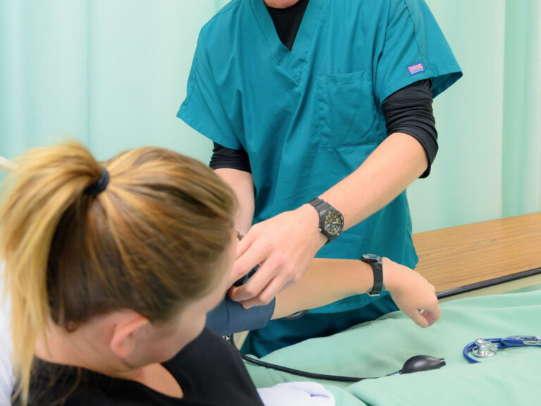
During an ultrasound scan, patients are usually asked to lie on a table while a technician or doctor applies a gel to the skin. The gel helps transmit sound waves effectively. The transducer is then moved gently over the targeted area to capture images. The procedure is comfortable, requires no recovery time, and patients can resume normal activities immediately afterward.
Conclusion
Ultrasound imaging is an indispensable diagnostic tool that helps detect, monitor, and manage a wide variety of internal organ and tissue conditions. Safe, accurate, and non-invasive, an Ultrasonogrāfija (USG) provides essential insights that guide medical care and promote early intervention. Whether for routine check-ups, pregnancy monitoring, or the investigation of internal health concerns, ultrasound remains one of the most trusted and effective methods for maintaining overall health and well-being.